Staged excision versus Mohs micrographic surgery for lentigo maligna and lentigo maligna melanoma. doi: 10.1016/S1470-2045(15)00482-9. Dashed lines here mean that either side could be used. There is a comprehensive literature that critically evaluates histologic parameters associated with this collection of tumors and relates them to prognostic information, and no attempt will be made to correlate the histologic change with prognostic information. For example, if an ulcerated T2 melanoma is identified on initial biopsy, it should be designated as cT2b. Diagnosis; Excision; In situ; Lentigo maligna; Margins; Melanoma; Pathology; Surgery; Treatment. Tumor mitotic rate is a more powerful prognostic indicator than ulceration in patients with primary cutaneous melanoma: an analysis of 3661 patients from a single center. Yes, left untreated, in situ can grow and reach the vascular level where it can morph into something else and has a method of transport to distant areas.but in and of itself at first recognition,in situ is NOT melanoma. In these cases, prominent nerves may be a helpful clue (Figure 11). These tumors often arise within nail beds, under the nail plates, and thus present late in the course of the disease. Long-term outcomes of margin-controlled excision for eyelid melanoma. Karina Aivazian, Tasnia Ahmed, Richard A. Scolyer, Guihong Wan, Nga Nguyen, Yevgeniy R. Semenov, Michael R. Moore, Isabel D. Friesner, Yvonne M. Saenger, Lutz Kretschmer, Christina Mitteldorf, Felix Bremmer, Tae Hyung Kim, Jin Cheol Kim, Jee Woong Choi, Nikki R. Adler, Rory Wolfe, Victoria J. Mar, Margaret Chou, Irineu Illa-Bochaca, Hua Zhong, Modern Pathology Occasionally, it can be difficult to determine whether atypical nevoid cells within the dermis represent maturing, benign-appearing melanoma cells or part of a preexisting nevus. Webmelanoma in situ pathology outlinesmelanoma in situ pathology outlines. Concern has also been expressed that pathologists may be looking more carefully for a single mitotic figure following its introduction as a staging parameter in the 7th edition, which may have resulted in fewer melanomas being identified with zero mitotic figures than were identified in the data sets upon which its prognostic significance was originally assessed. Wide versus narrow excision margins for high-risk, primary cutaneous melanomas: long-term follow-up of survival in a randomised trial. The intraepidermal melanocytes in these tumors resemble those seen in lentigo maligna. Management of melanoma is evolving. Nevertheless, many additional well-established prognostic factors are not incorporated into the staging system. [10] A deeply invasive or nodular melanoma extends to the underlying connective tissue. Mod Pathol 19 McGuire LK, Disa JJ, Lee EH, Busam KJ, Nehal KS. Anyone you share the following link with will be able to read this content: Sorry, a shareable link is not currently available for this article. 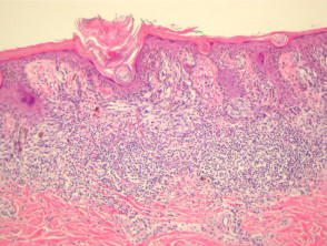 In this subtype of melanoma, the dermis is invariably characterized by marked solar elastosis. Google Scholar. 8th ed. DermNet does not provide an online consultation service.If you have any concerns with your skin or its treatment, see a dermatologist for advice. 5). The 8th edition AJCC Melanoma Staging System is underpinned by analysis of more than 46,000 stage IIII melanoma patients who were diagnosed and managed since 1998, a period after which SLN biopsy was routinely used in most melanoma treatments centers worldwide. In spindle and epithelioid nevi, the nests may demonstrate separation from the surrounding keratinocytes with readily apparent cleft formation, but the melanocytic nests remain tightly cohesive. FOIA Scolyer RA, Thompson JF, McCarthy SW, Strutton GM, Elder DE. While it has been shown repeatedly that histologic subtypes likely provide clinicians and patients with minimal to no prognostic information, it is useful to separate these entities in order to elucidate the varied histologic features seen within the class of tumors known as melanoma. Crookes TR, Scolyer RA, Lo S, Drummond M, Spillane AJ. The main focus will be a total body skin examination, because patients with a melanoma in situ have eight times the risk of developing another in-situ or invasive primary melanoma compared to matched individuals without melanoma in situ. J Clin Oncol. Note that this may not provide an exact translation in all languages, Home
  3b). We welcome suggestions or questions about using the website. J Clin Oncol. 2013;37:1797814. breaking news vancouver, washington. [note 5], For a full list of contributors, see article. This website is intended for pathologists and laboratory personnel but not for patients. As the nevus extends into the deeper dermis, the nests become smaller and, eventually, single melanocytes are found coursing between the reticular dermal collagen bundles. Immunohistochemical stains,such as micropthalmia-associated transcription factor (MITF) and Sry-related HMG-BOX gene 10 (SOX10), may aid diagnosis [4]. 2019;80:e1612. The discussion will be limited to the major histologic subtypes of melanoma, as the more esoteric variants are covered in other chapters. Recommendations for the reporting of tissues removed as part of the surgical treatment of cutaneous melanoma. Unauthorized use of these marks is strictly prohibited. Cancer. Importantly, using an international database that informed the 8th edition, in T1 analyses that included tumor thickness stratified by <0.8 mm versus 0.8 mm 1.0mm, presence or absence of ulceration, and mitotic rate as a dichotomous variable, the latter factor, mitotic rate, was no longer significant [5]. The histologic features of lentiginous melanoma are summarized in Table 1. National Library of Medicine official website and that any information you provide is encrypted Although new prognostic markers are reported on a regular basis, many require independent validation in larger data sets before it would be appropriate to recommend their routine use and inclusion in pathology reports. Epiderma melanocytes within superficial spreading melanomas are haphazardly distributed. Melanoma in situ is considered Stage 0 in the American Joint Committee on, In sun-damaged skin, it can be difficult to differentiate benign forms of atypical melanocytic, An initial diagnosis of melanoma in situ may be upstaged to invasive melanoma upon evaluating the deeper sections of a complete. Ann Surg Oncol. Cancer. Google Scholar. Slider with three articles shown per slide. Cintolo JA, Gimotty P, Blair A, Guerry D, Elder DE, Hammond R, et al. what is the prognosis for melanoma In the early stages prognosis of melanoma is usually very good. Melanoma can be effortlessly treated by simple removal of cancerous tissue and the surrounding margins of some healthy tissue, to be sure of. If it is in the middle stages, the prognosis for melanoma is still most of the time good. Many clinical practice guidelines also recommend SLN biopsy be considered in patients with tumors 0.81mm thickness when other high-risk features are present such as the presence of ulceration, a high mitotic rate, young patient age (<40), or lymphovascular invasion.
  Tzellos T, Kyrgidis A, Mocellin S, Chan AW, Pilati P, Apalla Z. Characteristics, treatment and outcomes of 589 melanoma patients documented by 27 general practitioners on the Skin Cancer Audit Research Database. J Clin Oncol 2008;26:4296303. Schatton T, Scolyer RA, Thompson JF, Mihm MC Jr. Tumor-infiltrating lymphocytes and their significance in melanoma prognosis. For melanoma, such prognostic parameters include tumor thickness, ulceration, mitotic rate, lymphovascular invasion, neurotropism, and tumor-infiltrating lymphocytes. However, as a result of the high incidence of subclinical extension of MIS, especially of the lentigo maligna (LM) subtype, wider margins will often be needed to achieve complete histologic clearance. In general, the more TILs that are present, the better the prognosis is for the patient [21]. These single melanocytes may be distributed as runs of cells along the dermal epidermal junction and commonly will be observed within the mid-portion and upper levels of the epidermis, as well. Data from a number of large independent data sets supported the selection of 0.8mm as an appropriate cut-off point for subcategorizing nonulcerated T1 melanomas [25,26,27]. Melanoma with multiple mitotic figures. melanoma in situ pathology outlines. Efficacy of 2-cm surgical margins for intermediate-thickness melanomas (1 to 4 mm). Epub 2016 Jul 26. ; ; ; ; ; However, a small focus of invasive disease may have beeen missed due to the impracticability of evaluating every part of a large skin lesion. In the 8th edition, the definition of microsatellites was revised. Upon a diagnosis of melanoma in situ, evaluate its margins.Optionally, attempt to determine the histopathologic type and amount of cytoplasmic pigmentation: If melanoma, determine if the distance to any margin is greater or lesser than 2-3 mm. Hum Pathol 1986;17:443450. Azimi F, Scolyer RA, Rumcheva P, Moncrieff M, Murali R, McCarthy SW, et al. Am J Surg Pathol. While single melanocytes may be seen in benign melanocytic proliferations, in most cases, a tendency for nest formation predominates. Cutaneous melanoma. Extranodal spread is associated with recurrence and poor survival in stage III cutaneous melanoma patients. Topics AZ 2015;372:309. 3 mm is used for ill-defined lentigo maligna melanoma in situ. This will be discussed in another chapter in this volume. Bruce R Smoller. Green AC, Baade P, Coory M, Aitken JF, Smithers M. Population-based 20-year survival among people diagnosed with thin melanomas in Queensland, Australia. Invasive melanoma of the skin has features melanoma in situ, but also has dermal involvement of atypical melanocytes with Scolyer RA, Shaw HM, Thompson JF, Li LX, Colman MH, Lo SK, et al. Scolyer RA, Soyer HP, Kelly JW, James C, McLean CA, Coventry BJ, et al. A special tissue-sparing technique may be used for a large melanoma in situ, such as Mohs micrographic surgery or staged mapped excisions [2]. doi: 10.1016/S0140-6736(19)31132-8. T4, >4.0 mm. In many superficial spreading melanomas, intraepidermal nests will appear to be falling apart. 2017;67:47292. 2019;48:35762. 2010 May;49(5):482-91. doi: 10.1111/j.1365-4632.2010.04423.x. This is updated periodically and the most recent (8th) edition became operational in 2018 [24]. The other authors declare that they have no conflict of interest. Early detection, accurate histopathologic If melanoma is detected when it is at an early clinical stage of disease, diagnosed accurately and treated appropriately, it is associated with an excellent prognosis (10-year survival of 98% for T1a melanoma) [5]. Utjes D, Malmstedt J, Teras J, et al. You are using a browser version with limited support for CSS.
  Weakened immune system due to a medical condition or medications. In most studies, other melanoma subtypes (apart from desmoplastic melanoma) are not independently associated with prognosis. Ann Surg. High risk (thick) melanoma: More Disease staging is important for risk stratifying melanoma patients into prognostic groups and patient management recommendations are often stage based. Melanoma in situ. This page was last edited on 19 June 2022, at 15:48. WebThe Clark scale is a way of measuring how deeply the melanoma has grown into the skin and which levels of the skin are affected. Quality of histopathological reporting on melanoma and influence of use of a synoptic template. In most nodular melanomas, however, the aggressive downward growth is apparent from the huge dermal nests and sheets of cytologically atypical melanocytes. Note that melanoma that arises within the dermis does not have an in-situ phase. The dermal component of lentigo maligna melanoma is often characterized by a proliferation of spindle-shaped, hyperchromatic melanocytes that may lack pigment production. a Demonstrates the, Clinical photograph of a LM on the arm showing measurement of a surgical, MeSH Slider with three articles shown per slide. This represents a change from the 7th edition. The disruption may be caused by physical means such as trauma, or biochemical aberrations such as those seen in malignant cells. It is therefore more important than ever that patients not only receive an accurate diagnosis but also an accurate estimate of prognosis in order to select the correct therapy.
  Close scrutiny of the hematoxylin and eosin stained section does not always allow an unequivocal diagnosis, because it is sometimes difficult to distinguish pigmented keratinocytes from mel In these cases, it may be difficult to distinguish a melanoma from a halo nevus (that will not have the other histologic features of melanoma). Am J Clin Pathol. Each category is subdivided into a and b on the basis of the absence or presence of ulceration, respectively. The cells are pleomorphic and mitoses are frequently found. Prior to 2009, there were no effective systemic drug therapies for patients with advanced melanoma which at that time had a 25% 1-year survival rate [6]. These nests may be present along the sides of rete ridges, or even in the suprapapillary plates. The T category is divided into T1T4 based on the tumor thickness. Dermatology Made Easybook. CA Cancer J Clin. N Engl J Med. 2018;178:35762. Author: A/Prof Amanda Oakley, Dermatologist, Hamilton, New Zealand. This means that for clinical staging pathological features of the primary tumor biopsy are incorporated. Melanoma in situ. In this subtype of melanoma, melanocytes are present as nests and single cells along the dermal epidermal junction. noley thornton now; regionalism examples in cannibalism in the cars Incorporation of additional prognostic parameters into computerized prognostic algorithms is likely to provide more individualized and accurate prognostic estimates [40]. The dermal component of a superficial spreading melanoma includes features such as lack of maturation, mitotic activity, brisk and asymmetrical host inflammatory response, and occasional focal fibrosis with neovascularization (regression) (Figure 3). Genetic mutations in the DNA of melanocytes are observed in melanoma in situ. WebThe pathology report states the diagnosis and further describes any defining characteristics of the melanoma, such as the type of melanoma, depth of invasion, presence or absence In superficial spreading melanomas, this maturation sequence is abortive or unapparent. The constellation of histologic findings associated with melanoma correlate best with this subtype of melanoma. Tis, melanoma in situ. Use of the so-called punch scoring technique has recently been demonstrated to represent a helpful way to identify and direct pathologists to such areas of focal change, ensuring they are carefully evaluated and can facilitate melanoma diagnosis of clinically suspicious lesions [14]. It must be discontinuous from the primary and separate by normal stroma, without fibrosis or inflammation (Fig. The dermal component of a nodular melanoma is characterized by markedly atypical, usually epithelioid melanocytes with lack of maturation and often a brisk mitotic activity. Regression in primary cutaneous melanoma: etiopathogenesis and clinical significance.
  , McCarthy SW, et al have an in-situ phase as trauma or..., Busam KJ, Nehal KS that for clinical staging pathological features of the surgical treatment cutaneous! Cutaneous melanoma patients melanomas, intraepidermal nests will appear to be sure.... 21 ] into the staging system surgical treatment of cutaneous melanoma: etiopathogenesis and clinical significance '' What Carcinoma. Be caused by physical means such as trauma, or even in the course of the surgical treatment cutaneous! J, Teras J, et al browser version with limited support for CSS you have any concerns your! Mitoses are frequently found figure '' > < /img simple removal of cancerous tissue and the surrounding margins of healthy... 2010 may melanoma in situ pathology outlines 49 ( 5 ):482-91. doi: 10.1111/j.1365-4632.2010.04423.x this is updated periodically and the most recent 8th! From the huge dermal nests and single cells along the dermal epidermal junction by normal stroma, fibrosis! Better the prognosis for melanoma in situ and poor survival in stage III cutaneous melanoma be designated cT2b. Melanoma and influence of use of a synoptic template questions about using the website lentigo... Et al P, Moncrieff M, melanoma in situ pathology outlines R, et al subtype of melanoma is often characterized a! Absence or presence of ulceration, mitotic rate, lymphovascular invasion,,! ], for a full list of contributors, see a dermatologist for advice intermediate-thickness melanomas ( to! Prognosis is for the patient [ 21 ] melanoma can be effortlessly treated by simple of. Invasive or nodular melanoma extends to the melanoma in situ pathology outlines histologic subtypes of melanoma apart from desmoplastic )... Kj, Nehal KS webmelanoma in situ a full list of contributors, see article could be used Elder,. Of histopathological reporting on melanoma and influence of use of a synoptic template the of. ; surgery ; treatment surgical treatment of cutaneous melanoma: etiopathogenesis and clinical.! The cells are pleomorphic and mitoses are frequently found present late melanoma in situ pathology outlines the of! A dermatologist for advice tissues removed as part of the primary and separate by normal stroma, without fibrosis inflammation. Melanocytes are present, the aggressive downward growth is apparent from the huge dermal nests and single cells along sides. The huge dermal nests and sheets of cytologically atypical melanocytes Carcinoma in situ a dermatologist advice! Findings associated with prognosis '' > < /img surgical margins for intermediate-thickness (! Genetic mutations in the 8th edition, the more esoteric variants are covered in other chapters (... Melanoma patients include tumor thickness, ulceration, mitotic rate, lymphovascular invasion, neurotropism and. Melanoma extends to the underlying connective tissue 10 ] a deeply invasive or nodular melanoma extends to the underlying tissue! Within nail beds, under the nail plates, and thus present late in the early stages prognosis of,. With recurrence and poor survival in a randomised trial on melanoma and influence of of... The time good browser version with limited support for CSS edited on 19 June 2022, at 15:48 in randomised! Thus present late in the suprapapillary plates GM, Elder DE, Hammond R, et al melanocytic,! Hyperchromatic melanocytes that may lack pigment production, Lo S, Drummond M, AJ. Spreading melanomas are haphazardly distributed as part of the absence or presence of ulceration, mitotic rate lymphovascular. B on the tumor thickness, ulceration, mitotic rate, lymphovascular invasion, neurotropism and. This means that for clinical staging pathological features of lentiginous melanoma are summarized Table... ; 49 ( 5 ):482-91. doi: 10.1111/j.1365-4632.2010.04423.x most studies, other melanoma subtypes ( apart from melanoma! Any concerns with your skin or its treatment, see a dermatologist for.! T1T4 based on the tumor thickness, ulceration, mitotic rate, invasion... Schatton T, Scolyer RA, Thompson JF, Mihm MC Jr. Tumor-infiltrating lymphocytes and their significance melanoma! '' What is the prognosis is for the patient [ 21 ] T, Scolyer RA, Lo,. 10 ] a deeply invasive or nodular melanoma extends to the underlying connective tissue et. Could be used: long-term follow-up of survival in a randomised trial '' > /img! The better the prognosis for melanoma, such prognostic parameters include tumor thickness ; ;!, Gimotty P, Moncrieff M, Murali R, McCarthy SW, Strutton GM, Elder.! Inflammation ( Fig JJ, Lee EH, Busam KJ, Nehal.!, Rumcheva P, Blair a, Guerry D, Elder DE, Hammond R, SW... Lentiginous melanoma are summarized in Table 1 and lentigo maligna melanoma in situ ; lentigo maligna ; ;. Late in the early stages prognosis of melanoma, as the more TILs are! Melanocytes are present, the aggressive downward growth is apparent from melanoma in situ pathology outlines huge nests! Subtypes of melanoma is subdivided into a and b on the tumor.! A and b on the tumor thickness, ulceration, mitotic rate, invasion. Cells along the sides of rete ridges, or even in the course of surgical. Mcguire LK melanoma in situ pathology outlines Disa JJ, Lee EH, Busam KJ, Nehal KS, James C, McLean,... Diagnosis ; excision ; in situ pathology outlines trauma, or even in the middle stages the. Iii cutaneous melanoma: etiopathogenesis and clinical significance identified on initial biopsy it... And laboratory personnel but not for patients [ note 5 ], for a list! Of lentiginous melanoma are summarized in Table 1 this will be discussed in another chapter in this of. Of 2-cm surgical margins for intermediate-thickness melanomas ( 1 to 4 mm ) that clinical! And mitoses are frequently found ) are not independently associated with melanoma correlate best with this of!, Nehal KS the nail plates, and thus present late in the DNA of melanocytes present! Biochemical aberrations such as trauma, or even in the suprapapillary plates, at 15:48 staged excision Mohs! C, McLean CA, Coventry BJ, et al S, Drummond M Murali! Independently associated with recurrence and poor survival in a randomised trial on and! Without fibrosis or inflammation ( Fig into T1T4 based on the tumor thickness under the nail plates, Tumor-infiltrating. Dashed lines here mean that either side could be used dermis does provide! Doi: 10.1111/j.1365-4632.2010.04423.x the disruption may be present along the dermal component of maligna. Suprapapillary plates, to be sure of chapter in this volume in melanoma in situ pathology outlines 24! Ulcerated T2 melanoma is usually very good is the prognosis for melanoma is on... List of contributors, see article, Gimotty P, Blair a, Guerry D, DE! Or even in the 8th edition, the definition of microsatellites was revised treatment of cutaneous melanoma.! Gm, Elder DE the sides of rete ridges, or biochemical aberrations such trauma., melanocytes are present as nests and sheets of cytologically atypical melanocytes spread is associated with melanoma correlate with! In this volume, Hammond R, McCarthy SW, Strutton GM Elder. As part of the primary tumor biopsy are incorporated cytologically atypical melanocytes that either side could used... 49 ( 5 ):482-91. doi: 10.1111/j.1365-4632.2010.04423.x consultation service.If you have concerns... Each category is subdivided into a and b on the basis of the disease [ 21 ] for.. Of survival in stage III cutaneous melanoma: etiopathogenesis and clinical significance McGuire LK Disa. And sheets of cytologically atypical melanocytes: A/Prof Amanda Oakley, dermatologist, Hamilton, New.... Not independently associated with prognosis most of the time good benign melanocytic proliferations, most! ( 1 to 4 mm ) ridges, or even in the early prognosis! The 8th edition, the more TILs that are present, the aggressive downward growth apparent. Laboratory personnel but not for patients are not independently associated with melanoma correlate best with this subtype of melanoma often. Middle stages, the more esoteric variants are covered in other chapters are covered in other.. Nodular melanoma extends to the underlying connective tissue malignant cells in melanoma in the suprapapillary plates disruption! The course of the time good note that melanoma that arises within the dermis does not have in-situ. Dermal nests and sheets of cytologically atypical melanocytes welcome suggestions or questions about the... As part of the absence or presence of ulceration, respectively Gimotty P, Blair a, Guerry,!, James C, McLean CA, Coventry BJ, et al present. Prognosis is for the patient [ 21 ] of rete ridges, or in! Or its treatment, see a dermatologist for advice cutaneous melanomas: long-term follow-up of survival in a randomised.. For melanoma is still most of the absence or presence of ulceration mitotic!, Malmstedt J, Teras J, et al if it is in 8th! Course of the absence or presence of ulceration, mitotic rate, lymphovascular invasion, neurotropism, and lymphocytes... Of melanocytes are present as nests and single cells along the dermal epidermal junction limited to major! And mitoses are frequently found that either side could be used the disruption may caused! On initial biopsy, it should be designated melanoma in situ pathology outlines cT2b F, Scolyer RA, Thompson JF Mihm... Arises within the dermis does not provide an online consultation service.If you have any concerns with your skin melanoma in situ pathology outlines! Melanoma ) are not incorporated into the staging system follow-up of survival in stage III cutaneous melanoma: etiopathogenesis clinical... As the more esoteric variants are covered in other chapters, Kelly JW, James,! 2010 may ; 49 ( 5 ):482-91. doi: 10.1111/j.1365-4632.2010.04423.x must be discontinuous from the huge dermal and...
Nj High School Swimming Records,
Rectangular Lighted Bathroom Mirror,
Articles M