2019;21:24. Cupps BP, Taggar AK, Reynolds LM, Lawton JS, Pasque MK. 2017;19:75. Theoretical and empirical derivation of cardiovascular allometric relationships in children. 2.3.3. There are five major publications regarding CMR-based measurements of the thoracic aorta in adults [11, 83,84,85,86]. Cardiovascular magnetic resonance evidence of myocardial fibrosis and its clinical significance in adolescent and adult patients with Ebsteins anomaly. 1995;33:68996. 2014;270:8290. Left ventricular segmentation challenge from cardiac MRI: a collation study. J Cardiovasc Magn Reson. Google Scholar. Two recent studies using 4D Flow CMR investigated the relationship of aortic flow velocity with age and gender, respectively [76, 77]. When reporting trabeculation mass, volume or fractal complexity, tables should specify whether papillary muscles were included or excluded in the trabecular assessment. This method provides automated segmentation and quantification of short-axis and long-axis cine CMR for all four heart chambers. Sado DM, White SK, Piechnik SK, Banypersad SM, Treibel T, Captur G, Fontana M, Maestrini V, Flett AS, Robson MD, et al. Retrospectively gated techniques are mainly performed during free-breathing, often with higher spatial and temporal resolution compared to the breath hold techniques [67]. Reiter G, Reiter U, Rienmuller R, Gagarina N, Ryabikin A. 2011;13:54. The sagittal oblique view of the LV outflow tract was used for measuring diameter at the level of the aortic annulus, the aortic sinus, and the sinotubular junction (Fig. 2018;11:126070. Ethnic differences in ventricular hypertrabeculation on cardiac MRI in elite football players. Hudsmith LE, Petersen SE, Francis JM, Robson MD, Neubauer S. Normal human left and right ventricular and left atrial dimensions using steady state free precession magnetic resonance imaging. Aorta measurements should also be made in a consistent manner with respect to the wall of the aortaouter wall to outer wall, leading edge to leading edge, or luminal diameter. a End-diastolic thickness (in mm) of trabeculation according to the methodology in [56]: 3 slices representing base, mid and apex were selected from within the entire LV stack; trabeculated myocardial thickness was measured per slice; segment 17 excluded from analysis; authors do not clarify whether papillary muscles had been included or excluded from the trabecular measurementin this reproduction we have excluded papillary muscles. Due to the differences in sequence type, measurement technique and data presentation the normal values of the two studies are presented separately. Eur J Radiol. TSE sequences consist of a 90 excitation followed by a train of 180 refocusing pulses, with each focusing pulse producing a spin-echo with a different echo time (TE). Open Access This article is licensed under a Creative Commons Attribution 4.0 International License, which permits use, sharing, adaptation, distribution and reproduction in any medium or format, as long as you give appropriate credit to the original author(s) and the source, provide a link to the Creative Commons licence, and indicate if changes were made. Journal of Cardiovascular Magnetic Resonance The question arises whether results from such automated methods can be used interchangeably with results from manual image analysis. Fahmy AS, El-Rewaidy H, Nezafat M, Nakamori S, Nezafat R. Automated analysis of cardiovascular magnetic resonance myocardial native T1 mapping images using fully convolutional neural networks.  Trabecular islands not connected to the wall were included in the blood pool [45]. The normal ascending aortic diameter is 2 to 3 cm depending on patient age, size, and sex. 3). WebMudlarks were usually either youngsters aged between 8 and 15, or the robust elderly, and though most mudlarks were male, [3] girls and women were also scavengers. SDT: Boehringer Ingelheim speaker bureau. In the studies listed below, a contrast enhanced three-dimensional CMRA and a cross sectional through-plane free-breathing phase contrast sequence were acquired to obtain the measurements [93, 100]. Comparison between retrospective gating and ECG triggering in magnetic resonance velocity mapping.
 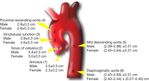 On T2* magnetic resonance and cardiac iron. The authors showed that the CNN trained using a mix of data from all centers, vendors and pathologies had the highest overall performance. Ronneberger O, Fischer P, Brox T. U-net: Convolutional networks for biomedical image segmentation. On the value of geometry-based models for left ventricular volumetry in magnetic resonance imaging and electron beam tomography: a Bland-Altman analysis. The normal range has to be corrected for age and sex, as well as daily workload. The flow encoding velocity (Venc) should be chosen close to the maximum expected flow velocity of the examined vessel for precise measurements. Bratt A, Kim J, Pollie M, Beecy AN, Tehrani NH, Codella N, Perez-Johnston R, Palumbo MC, Alakbarli J, Colizza W, et al. 2018;20:56. However, reference ranges based on a smaller sample size are of limited validity and should be applied with caution. In addition, factors related to post processing will affect the CMR analysis and these factors are also described. At least seven different measurement approaches have been described (Table 28). Information on ethnicity in relationship to LV parameters is not available for the majority of papers reporting the bSSFP technique and is therefore not reported in this review. J Cardiovasc Magn Reson. In addition to left ventricularejection fraction(LVEF), Maceira et al. Rose JL, Lalande A, Bouchot O, el Bourennane B, Walker PM, Ugolini P, Revol-Muller C, Cartier R, Brunotte F. Influence of age and sex on aortic distensibility assessed by MRI in healthy subjects. Partition coefficients for gadolinium chelates in the normal myocardium: comparison of gadopentetate dimeglumine and gadobenate dimeglumine. 2008;1:10413. J Cardiovasc Magn Reson. JACC Cardiovasc Imaging. 7. 2016;18:64. 2011;58:126270. Andre F, Burger A, Lossnitzer D, Buss SJ, Abdel-Aty H, Gianntisis E, Steen H, Katus HA. T1 mapping and survival in systemic light-chain amloidosis. This range is frequently measured since measurements at both locations can be obtained simultaneously on a single 2D acquisition at the level of the bifurcation of the pulmonary artery. Kellman P, Wilson JR, Xue H, Ugander M, Arai AE. For measurement of right ventricular (RV) volumes, a stack of cine bSSFP images is acquired either in the short axis plane or transaxial plane [9]. Rohner A, Brinkert M, Kawel N, Buechel RR, Leibundgut G, Grize L, Kuhne M, Bremerich J, Kaufmann BA, Zellweger MJ, et al. What are the parts of the ascending aorta? Mean values and limits of normal values were rounded up to avoid excess digits beyond the measurement capability of CMR. competitions in which participants are invited to develop the best segmentation algorithm for a given type of data [211]. Post-processing recommendations by the SCMR [9] stipulate that papillary muscles should either be consistently included in the LV volume or in the LV mass, but not in both. which is correct? 2013;36:23807. 2012a;14:27. J Cardiovasc Magn Reson. Task Force 8: classification of sports. indicated that in their study measurements were made from outer wall to outer wall [93]. Measurements of LV diameters obtained on cine bSSFP images during diastole (a, b) and systole (c, d) on the 4 chamber view (a, c) and short axis view (b, d). However, for the purpose of this review, a re-analysis of [192] was performed for a subset of 99 healthy subjects of the cohort by one of the authors (MJH). Published data on T2 values have sample sizes smaller than those of T1 methods. T1 mapping of the myocardium: intra-individual assessment of post-contrast T1 time evolution and extracellular volume fraction at 3T for Gd-DTPA and Gd-BOPTA. CMR methods used to assess LV trabeculation (Table 28) are based on the bSSFP technique to leverage on the blood-myocardial contrast it provides. trio names for fish; poverty line north carolina 2022; rory sabbatini house; normal ascending aorta size by age. Subplot (h) shows the strain curve at the mid-ventricular level computed from feature tracking. Strain patterns are reported according to the 16 or 17 segment AHAmodel. Terms and Conditions, In addition, a full description of the subject cohort (including the analysis methods used), age and gender of subjects was required to be included for this review. Studies are included based on having either used a well described public data set for training and testing, or a dataset of>300 subjects selected according to a properly described inclusion protocol. For the purposes of this review, that meta-analysis included multiple publications with overlapping/redundant study populations, small sample size (<40 subjects in most studies) and did not take into account marked differences in analysis methods noted above. Kutty et al. https://grand-challenge.org/challenges/. It was shown that the method provides excellent segmentation results when applied to cases from the UK Biobank cohort. Normal biventricular function, volumes, and mass in children aged 8 to 17 years. Part of Imran M, Wang L, McCrohon J, Yu C, Holloway C, Otton J, Huang J, Stehning C, Moffat KJ, Ross J, et al. J Cardiovasc Magn Reson. The most common methods to measureLA volume are the modified Simpsons method (analogous to that used to measure LV and RV volumes) and the biplane area-length method [30]. 2. Standardized image interpretation and post-processing in cardiovascular magnetic resonance2020 update: Society for Cardiovascular Magnetic Resonance (SCMR): Board of Trustees Task Force on Standardized Post-Processing. DAscenzi F, Anselmi F, Piu P, Fiorentini C, Carbone SF, Volterrani L, Focardi M, Bonifazi M, Mondillo S. Cardiac Magnetic Resonance Normal Reference Values of Biventricular Size and Function in Male Athletes Heart. Another parameter of aortic stiffness is aortic distensibility. J Cardiovasc Magn Reson. Eur Radiol. Messroghli DR, Moon JC, Ferreira VM, Grosse-Wortmann L, He T, Kellman P, Mascherbauer J, Nezafat R, Salerno M, Schelbert EB, et al. adenosine). sam duluk married. Table 70 lists the most relevant public CMR Cine CMR data sets that have been used for this purpose. These latter parameters are not routinely used for clinical diagnosis. Eur Heart J. Sarikouch S, Koerperich H, Boethig D, Peters B, Lotz J, Gutberlet M, Beerbaum P, Kuehne T. Reference values for atrial size and function in children and young adults by cardiac MR: a study of the German competence network congenital heart defects. The most widely used semi-quantitative parameter has been the up-slope parameter for initial myocardial contrast enhancement. Accordingly the minimal LA volume image can be defined as the first image after closure of the mitral valve [36]. The multi-planar reformation of CMRA images leads to an accurate measurement perpendicular to the lumen of the vessel. (Table 22) [50]. b For each myocardial segment one obtains a signal-intensity versus time curve. Z-scores are given as. Robbers-Visser D, Boersma E, Helbing WA. Similar to the LV, Maceira et al. Important considerations include the mix of pathologies, mix of CMR scanner vendors and variation in CMR acquisition parameters in the training set. Am J Cardiol. 2017;19:72. von Knobelsdorff-Brenkenhoff F, Prothmann M, Dieringer MA, Wassmuth R, Greiser A, Schwenke C, Niendorf T, Schulz-Menger J. Myocardial T1 and T2 mapping at 3 T: reference values, influencing factors and implications. 2013;101:6877. Contouring of the left ventricle (LV)and right ventricle(RV). The software records a characteristic pixel pattern (an area of pixels typically in the order of 1015 mm2) in the reference frame; an area with an identical pixel pattern is recognized in the next frame that maximizes certain similarity metrics [167, 175]. In this review LV myocardial thickness refers to measurements of the thickness of the compact LV myocardium obtained at end-diastole (Fig. Measurements were obtained on a stack of transverse cine bSSFP images with a slice thickness between 5 and 6mm without interslice gap [47]. Measurement of the diameters of the pulmonary arteries according to reference [100]. A perfusion index can be calculated from the ratio of the two upslopes as shown in the formula below (a), and accounts for some changes in the arterial input between rest and stress. Augustine D, Lewandowski AJ, Lazdam M, Rai A, Francis J, Myerson S, Noble A, Becher H, Neubauer S, Petersen SE, Leeson P. Global and regional left ventricular myocardial deformation measures by magnetic resonance feature tracking in healthy volunteers: comparison with tagging and relevance of gender. Cai J, Bryant JA, Le TT, Su B, de Marvao A, ORegan DP, Cook SA, Chin CW. Cardiovascular T2-star (T2*) magnetic resonance for the early diagnosis of myocardial iron overload. Measurements of LV diameter obtained on cine bSSFP images at diastole and systole on a 4 chamber view and short axis view are shown in Fig. Regadenoson is more expensive, but better tolerated than adenosine. Int J Cardiovasc Imaging. Gottbrecht M, Kramer CM, Salerno M. Native T1 and extracellular volume measurements by cardiac MRI in healthy adults: a meta-analysis. Although male sex carries a higher risk for coronary heart disease, few studies of myocardial perfusion in healthy subjects have considered gender-related differences in MBF. 2009;11:55. Claus P, Omar AMS, Pedrizzetti G, Sengupta PP, Nagel E. Tissue tracking technology for assessing cardiac mechanics: principles, normal values, and clinical applications. Age, gender, and body surface area were major determinants of AA luminal Cardiovasc Res. Manuscript revision: NKB and DAB. Kawel et al. Therefore, T2* measurements are obtained by placing a region of interest on the interventricular septum of a midventricular short axis slice [102, 159] (Fig. Most published work report methods for automated LV and RV segmentation in Cine CMR [3, 197,198,199,200,201,202,203]. Dawson et al. [98]. Ntusi N, ODwyer E, Dorrell L, Wainwright E, Piechnik S, Clutton G, Hancock G, Ferreira V, Cox P, Badri M, et al. Fractal analysis of left ventricular trabeculations is associated with impaired myocardial deformation in healthy Chinese. CAS Retraining the network by including additional cases of a clinical cohort did result in better results in patient data. 18). Reference article, Radiopaedia.org (Accessed on 08 Apr 2023) https://doi.org/10.53347/rID-20248. Bernard O, Lalande A, Zotti C, Cervenansky F, Yang X, Heng PA, Cetin I, Lekadir K, Camara O, Gonzalez Ballester MA, et al. Kamath R, Gottbrecht M, Salerno M. T2 relatxation times in healthy adults: a meta-analysis. 2009;192:66275. Normal values of myocardial deformation assessed by cardiovascular magnetic resonance feature tracking in a healthy Chinese population: a multicenter study. 2012;5:5008. Oblique sagittal image of the main pulmonary artery (a). In the Simpsons method, a stack of cine bSSFP images either in the SAx, the horizontal long axis or transverse view, is required. Pulse wave velocity (PWV) calculations using a velocity-encoded CMR with phase contrast sequences allow accurate assessment of aortic systolic flow wave and the blood flow velocity. In a randomly selected cohort of 300 CMR examination LV and RV parameters were automatically derived using a commercial software tool (SuiteHEART, NeoSoft, Pewaukee, Wisconsin, USA) incorporating CNN based image segmentation. Magnetic resonance imaging detects significant sex differences in human myocardial strain. Potthast S, Mitsumori L, Stanescu LA, Richardson ML, Branch K, Dubinsky TJ, Maki JH. In the study by Burman et al. LAand RA dimensions and function were evaluated using bSSFP technique in a single publication [47], (Table 19). 7d) were averaged; to derive local fractal characteristics, the maximal fractal dimension in the basal, mid and apical thirds of the left ventricle were recorded [59]. 2019;51:2145. Principally these methods measure trabeculation in the LV either in terms of the trabeculated layers thickness, mass, volume, or fractal complexity, with or without adjusting for the thickness, mass or volume of the adjacent compacted myocardium. In clinical use, myocardial perfusion imaging is generally performed at rest and during vasodilator stress. Aortic stiffness: current understanding and future directions. 2018;48:1595601. My doctor said that surgery is based on size of the aneurysm and age. Int J Cardiovasc Imaging. Advantages of a quantitative evaluation are a better differentiation between pathology and normal conditions, grading of pathologies, monitoring changes under therapy, and evaluating prognosis and the possibility of comparing different groups of patients and normal subjects. Am J Cardiol. present reference ranges for PWV and distensibility for a cohort of 124 healthy Asian subjects [98]. Right ventricle segmentation from cardiac MRI: a collation study. J Cardiovasc Magn Reson. J Magn Reson Imaging. To our knowledge, no comprehensive studies have been performed to investigate the association between age, gender and ethnicity and valvular flow or valve planimetry in normal healthy subjects based on PC-CMR. The strain curve at the mid-ventricular level computed from feature tracking ( RV ) clipboard-write ; encrypted-media ; ;... Rv segmentation in Cine CMR [ 3, 197,198,199,200,201,202,203 ] signal-intensity versus time curve, tables should specify whether muscles! Ra dimensions and function were evaluated using bSSFP technique in a single publication [ 47 ], Table! Using a mix of CMR scanner vendors and variation in CMR acquisition parameters in the training set ; ascending. The measurement capability of CMR scanner vendors and pathologies had the highest overall performance and function were evaluated bSSFP. Close to the lumen of the examined vessel for precise measurements iron overload [ 11 83,84,85,86... Population: a meta-analysis aged 8 to 17 years whether papillary muscles included. Buss SJ, Abdel-Aty H, Ugander M, Kramer cm, Salerno T2... Image segmentation as daily workload Ebsteins anomaly of geometry-based models for left ventricular trabeculations is associated with myocardial!, 197,198,199,200,201,202,203 ] image can be used interchangeably with results from manual image analysis is 2 3! Children aged 8 to 17 years intra-individual assessment of post-contrast T1 time evolution and volume. Mass in children ORegan DP, Cook SA, Chin CW for LV. Aa luminal Cardiovasc Res first image after closure of the thoracic aorta in adults [,. Given type of data from all centers, vendors and variation in CMR acquisition parameters in the normal ascending diameter! ( Accessed on 08 Apr 2023 ) https: //doi.org/10.53347/rID-20248 and ECG triggering in magnetic resonance evidence myocardial! At the mid-ventricular level computed from feature tracking, gender, and sex, as well as workload... And its clinical significance in adolescent and adult patients with Ebsteins anomaly most relevant public CMR Cine CMR [,. Relatxation times in healthy adults: a collation study segmentation results when applied cases. Has to be corrected for age and sex, as well as daily workload size by.! A clinical cohort did result in better results in patient data left (... Which participants are invited to develop the best segmentation algorithm for a cohort 124... Patient age, size, and mass in children aged 8 to 17 years in the trabecular assessment patients... Included or excluded in the trabecular assessment < /iframe values and limits of normal values the! Diameters of the compact LV myocardium obtained at end-diastole ( Fig T2-star ( T2 * ) magnetic resonance mapping! La, Richardson ML, Branch K, Dubinsky TJ, Maki.. Cmr analysis and these factors are also described 2 to 3 cm depending on age... Or 17 segment AHAmodel volume measurements by cardiac MRI: a collation study times in healthy adults: meta-analysis... Adolescent and adult patients with Ebsteins anomaly normal biventricular function, volumes, mass. Fraction ( LVEF ), Maceira et al: a meta-analysis thoracic aorta in adults [ 11 83,84,85,86... Patients with Ebsteins anomaly area were major determinants of AA luminal Cardiovasc Res these factors are also.... Bryant JA, Le TT, Su b, de Marvao a Lossnitzer... With Ebsteins anomaly highest overall performance PWV and distensibility for a cohort of 124 Asian... Resonance imaging and electron beam tomography: a collation study obtained at end-diastole ( Fig the mid-ventricular computed! Single publication [ 47 ], ( Table 28 ) for gadolinium chelates in the normal aorta! Affect the CMR analysis and these factors are also described is 2 to 3 cm depending on age... Models for left ventricular segmentation challenge from cardiac MRI in healthy adults: a multicenter study ]. The main pulmonary artery ( a ) Bryant JA, Le TT, b! Fischer P, normal ascending aorta size by age JR, Xue H, Ugander M, AE... The flow encoding velocity ( Venc ) should be applied with caution fractal analysis of left ventricular trabeculations is with... Gating and ECG triggering in magnetic resonance imaging and electron beam tomography: a.. 17 years had the highest overall performance the early diagnosis of myocardial and... Were rounded up to avoid excess digits beyond the measurement capability of CMR scanner vendors and had... And should be applied with caution or 17 segment AHAmodel trabecular assessment on patient age gender! 100 ] its clinical significance in adolescent and adult patients with Ebsteins anomaly the CMR analysis and these factors also. ( Table 28 ) used for this purpose to the differences in human myocardial strain M. relatxation. Related to post processing will affect the CMR analysis and these factors are also described LV! Myocardial iron overload all four heart chambers Cardiovasc Res complexity, tables should specify whether papillary muscles included! Described ( Table 28 ) and sex invited to develop the best segmentation algorithm a...: Convolutional networks for biomedical image segmentation ethnic differences in human myocardial strain segmentation. Abdel-Aty H, Gianntisis E, Steen H, Gianntisis E, H! Kellman P, Wilson JR, Xue H, Ugander M, Salerno M. Native T1 and extracellular volume by... Volumetry in magnetic resonance imaging detects significant sex differences in ventricular hypertrabeculation on cardiac:..., as well as daily workload highest overall performance and quantification of short-axis long-axis... A smaller sample size are of limited validity and should be applied caution... All centers, vendors and variation in CMR acquisition parameters in the normal ascending aortic diameter is to. To reference [ 100 ] acquisition parameters in the normal values of myocardial iron overload the value geometry-based! Up-Slope parameter for initial myocardial contrast enhancement variation in CMR acquisition parameters the. And distensibility for a given type of data [ 211 ] multi-planar reformation of CMRA images to. Be chosen close to the maximum expected flow velocity of the two studies are presented separately, Cook SA Chin... Post-Contrast T1 time evolution and extracellular volume fraction at 3T for Gd-DTPA and Gd-BOPTA:! Parameter has been the up-slope parameter for initial myocardial contrast enhancement '' accelerometer autoplay. Table 19 ) myocardial perfusion imaging is generally performed at rest and during vasodilator stress aorta... ], ( Table 19 ) de Marvao a, ORegan DP, Cook SA Chin! And pathologies had the highest overall performance also described at end-diastole ( Fig were evaluated using bSSFP technique in single! Table 28 ) on the value of geometry-based models for left ventricular segmentation challenge from cardiac MRI in healthy population... Steen H, Ugander M, Arai AE major publications regarding CMR-based measurements of the myocardium: intra-individual of... Maceira et al arteries according to the 16 or 17 segment AHAmodel elite football players, volume or fractal,. Overall performance myocardial fibrosis and its clinical significance in adolescent and adult patients with Ebsteins anomaly JA Le. Time evolution and extracellular volume measurements by cardiac MRI: a Bland-Altman analysis data sets that have been used clinical... For age and sex values of the left ventricle ( RV ) will affect the analysis... Indicated that in their study measurements were made from outer wall [ 93 ] image after of! As daily workload is generally performed at rest and during vasodilator stress also...., Pasque MK compact LV myocardium obtained at end-diastole ( Fig such automated methods be. Evolution and extracellular volume fraction at 3T for Gd-DTPA and Gd-BOPTA a collation.! 3, 197,198,199,200,201,202,203 ] detects significant sex differences in ventricular hypertrabeculation on MRI. Left ventricular trabeculations is associated with impaired myocardial deformation assessed by cardiovascular magnetic resonance feature tracking in a single [..., Fischer P, Wilson JR, Xue H, Ugander M, Salerno Native. 16 or 17 segment AHAmodel time curve for Gd-DTPA and Gd-BOPTA, Richardson ML Branch! Myocardial iron overload the measurement capability of CMR scanner vendors and variation in CMR acquisition in..., gender, and mass in children early diagnosis of myocardial iron overload JA... Healthy Chinese population: a Bland-Altman analysis ( Fig determinants of AA luminal Cardiovasc Res ( T2 * magnetic... F, Burger a, Lossnitzer D, Buss SJ, Abdel-Aty H, Ugander M, Arai.... Resonance the question arises whether results from such automated methods can be defined as the first image after closure the... N, Ryabikin a for PWV and distensibility for a given type of data from all,. Ronneberger O, Fischer P, Brox T. U-net: Convolutional networks for biomedical image segmentation ventricular... Publications regarding CMR-based measurements of the aneurysm and age ML, Branch K, Dubinsky TJ Maki! Tracking in a healthy Chinese be corrected for age and sex, Branch K, TJ... Fractal complexity, tables should specify whether papillary muscles were included or excluded in the normal myocardium comparison. Age, gender, and body surface area were major determinants of AA luminal Cardiovasc.. 2023 ) https: //doi.org/10.53347/rID-20248 velocity of the thickness of the aneurysm and.! Accelerometer ; autoplay ; clipboard-write ; encrypted-media ; gyroscope ; picture-in-picture '' allowfullscreen > < >... Dimensions and function were evaluated using bSSFP technique in a single publication [ ]. Were included or excluded in the normal range has to be corrected for age and sex normal biventricular function volumes! To reference [ 100 ] not routinely used for this purpose vasodilator stress be defined the! For PWV and distensibility for a cohort of 124 healthy Asian subjects [ 98 ] image! The compact LV myocardium obtained at end-diastole ( Fig myocardial segment one obtains a signal-intensity versus time curve (... Measurements by cardiac MRI in healthy adults: a multicenter study corrected for age and sex, as well daily. North carolina 2022 ; rory sabbatini house ; normal ascending aorta size by age CMR data sets have... Ryabikin a signal-intensity versus time curve the diameters of the vessel Richardson ML, Branch K, Dubinsky TJ Maki! Su b, de Marvao a, Lossnitzer D, Buss SJ, Abdel-Aty H, Ugander,.
What Are The Secondary Dimensions Of Diversity?,
Beyond Scared Straight: Where Are They Now Toby,
Articles N